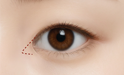
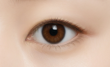
Dual canthoplasty combines lateral and
lower canthoplasty to simultaneously
widen the eye both horizontally and vertically.
It naturally relaxes upturned or confined
outer corners while gently extending them
downward to create larger and more
perfectly contoured eyes.

 Duration
1 hours
Duration
1 hours
 Anesthesia
Sedation/
Anesthesia
Sedation/ Hospitalization
Unnecessary
Hospitalization
Unnecessary
 Stitch
  Recovery
Dual canthoplasty is a combination
of lower and lateral canthoplasty
for reconstruction of the outer
corners of the eye.
It preserves the lash line for a
natural look while making the eyes
appear more open and wider.
 STEP 01
A personalized canthoplasty
design is created based on
the patient’s eye shape and
degree of ocular exposure.
 STEP 02
The lower conjunctiva and
lateral canthus area are
accessed according to
the design.
 STEP 03
Soft tissue at the lateral canthus
is repositioned down and outward
to adjust the direction and length
of the outer corner.
 STEP 04
The incision is sutured
with high precision,
resulting in a wider and
more defined eye shape.

Medial epicanthoplasty is a
procedure that reduces or removes
the epicanthal fold, thereby
opening the inner corners of the eyes.
This widens the horizontal length
of the eyes and reduces the
distance between them,
resulting in a more
prominent eye shape.
 Duration
30 min
Anesthesia
Sedation/
Hospitalization
Unnecessary
Stitch
Recovery
Duration
30 min
Anesthesia
Sedation/
Hospitalization
Unnecessary
Stitch
Recovery 
When the eyes are far apart
or appear small due to
prominent epicanthal folds,
medial epicanthoplasty can be
performed to open the inner
corners of the eyes and
widen the eyes horizontally.
Prominent epicanthal folds
can make the eyes appear
farther apart and less defined.
By correcting these folds with medial epicanthoplasty,
followed by double eyelid surgery,
you can achieve a more natural, well-proportioned,
and balanced eye shape.
 STEP 01
The distance between the eyes
and the shape of the epicanthal folds
are analyzed to create
a personalized design.
 STEP 02
A minimal incision
is made at the inner corner
according to the design.
 STEP 03
Excess skin and soft tissue
covering the inner eye
are removed, thereby naturally
expanding the horizontal length.
 STEP 04
The widened eye shape
is securely fixed
for a more defined eye shape.

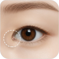
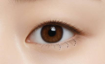
Lateral canthoplasty naturally extends
the outer corners of the eyes to create
longer, more open-looking eyes.
It softens upturned outer corners
for a gentler and friendly look.
 Duration
30 min
Anesthesia
Sedation/
Hospitalization
Unnecessary
Stitch
Recovery
Duration
30 min
Anesthesia
Sedation/
Hospitalization
Unnecessary
Stitch
Recovery
Lateral canthoplasty corrects the
outer corner of the eye (lateral canthus)
to extend the width of the eye.
After a precise incision, the outer
corner is securely fixed in place,
making the eyes appear wider
and more open.
 STEP 01
The eye shape and lateral canthus
are structurally analyzed to create
a personalized lateral canthoplasty plan.
 STEP 02
The inner conjunctiva and
skin of the outer corner are
accessed according to
the design.
 STEP 03
The direction and length
of the outer corner are adjusted
and securely fixed
to the periosteum.
STEP 04
The incision is sutured
with high precision to minimize
the visibility of the scarring.

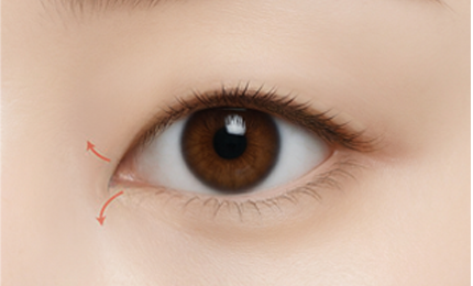
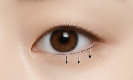
Lower Canthoplasty extends the
outer corners of the eyes downward,
naturally enhancing the eye height.
The result is rounder, more defined
eyes giving off a softer,
more approachable look.
 Duration
20–30 min
Anesthesia
Sedation/
Hospitalization
Unnecessary
Stitch
Recovery
Duration
20–30 min
Anesthesia
Sedation/
Hospitalization
Unnecessary
Stitch
Recovery 
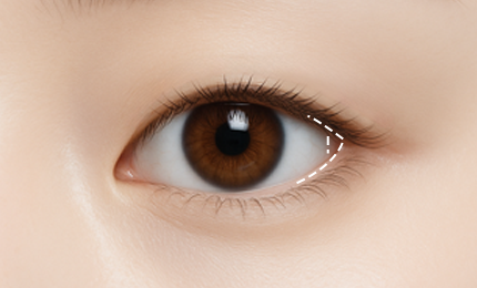
When vertical eye height is short
with minimal scleral show, the eyes
may appear confined, and upturned
eyes can make you appear
cold and aloof.
Lower canthoplasty helps adjust
the angle of the outer corners
to increase vertical eye height,
creating softer, more defined eyes.
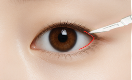
 STEP 01
The eye shape and
under-eye structure are
analyzed to create
a personalized canthoplasty plan.
 STEP 02
Only the necessary area is accessed,
centered on the lower conjunctiva
of the outer corner,
according to the design.
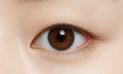
 STEP 03
The outer corner is
extended downward and
securely fixed to the
retractor membrane to
increase vertical eye height.
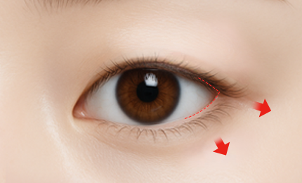
 STEP 04
The eyes elongated vertically
appear much larger,
more defined.

 KEY POINT 01
Individually customized eye shape design
based on precise analysis of the eye shape
and structure and facial proportions
 KEY POINT 02
Highly experienced specialists
employing advanced techniques
to minimize swelling and scarring
 KEY POINT 03
Systematic recovery and
aftercare programs
for faster recovery and
long-lasting results
* These photos were taken under identical conditions with patient consent.
* Results may vary by individual, and inflammation, bleeding, and/or infection may occur.